

سلامت گوش
Otoscopy vs Tympanometry vs Audiometry: What Does Each Test Check?
Quick answer
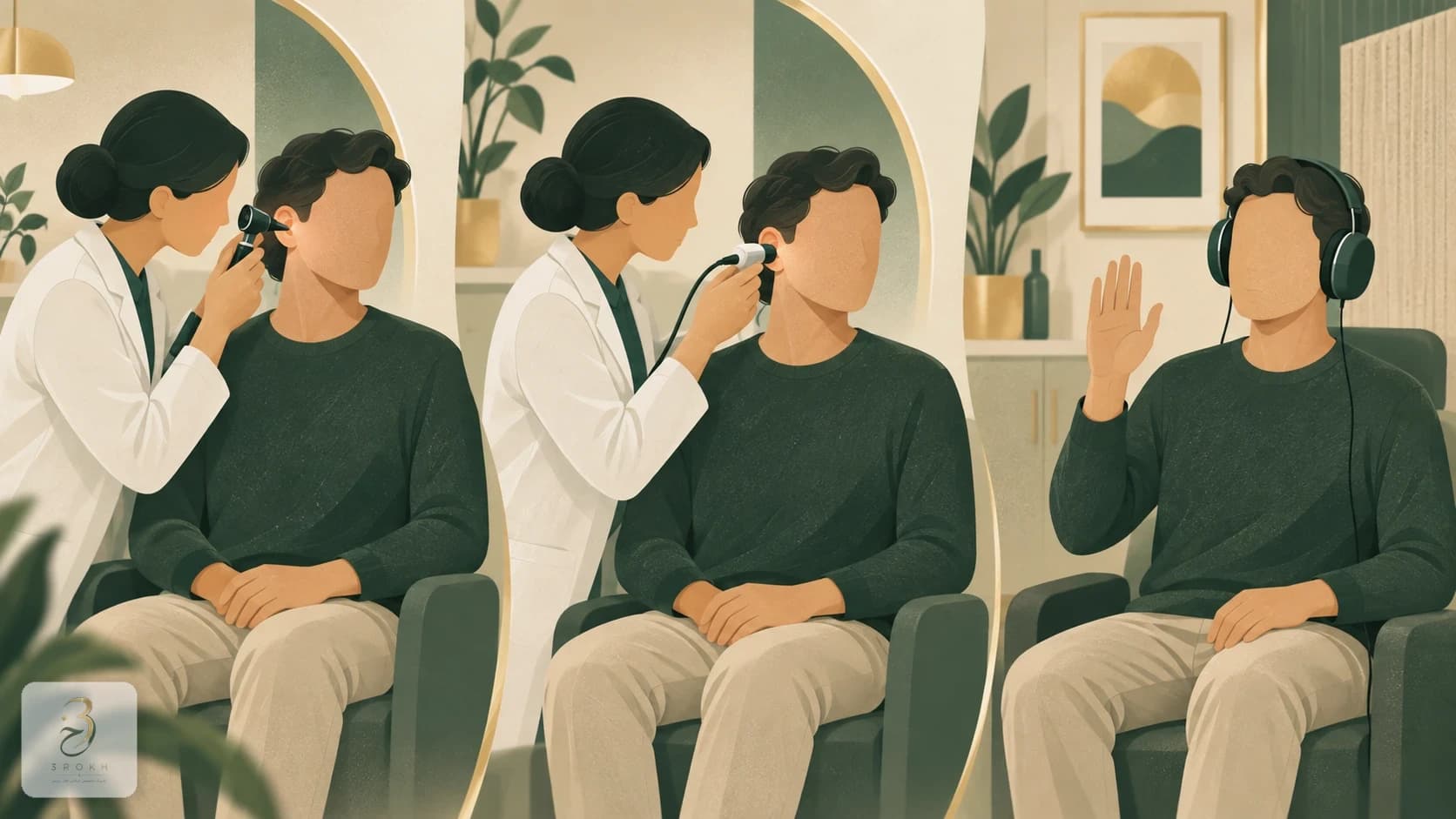
Otoscopy views the ear canal and eardrum, tympanometry measures middle-ear mechanics and pressure response, and audiometry measures hearing thresholds. The tests complement one another, so a normal result on one does not replace the others.
- Otoscopy
- Visual canal and eardrum examination
- Tympanometry
- Middle-ear mechanical function
- Audiometry
- Hearing thresholds and pattern
- Service page
- /services/otoscopy
Reliable interpretation combines symptoms, examination and test results.
Key points
- Otoscopy examines the visible ear canal and eardrum.
- Tympanometry measures how the eardrum and middle ear respond to pressure changes.
- Audiometry measures hearing thresholds across different frequencies.
- A normal result on one test does not prove that the other two are normal.
Short answer: these tests are not interchangeable
Otoscopy checks the visible structures of the ear canal and eardrum. Tympanometry assesses how the eardrum and middle-ear system respond to pressure. Audiometry measures the quietest sounds a person can hear across frequencies. A normal result on one test therefore does not prove that the other two areas are normal.
The tests answer different questions: what can be seen, how the middle ear transmits sound mechanically, and what the person can actually hear. ASHA lists otoscopy, acoustic immittance and audiometry as separate components of a comprehensive hearing assessment.
At-a-glance comparison
| Test | Main question | What it assesses | Typical output | Main limitation |
|---|---|---|---|---|
| Otoscopy | What can be seen inside the ear? | Ear canal and visible eardrum | Visual description | Does not measure hearing thresholds |
| Tympanometry | How does the middle ear respond to pressure? | Eardrum mobility, middle-ear pressure and mechanics | Tympanogram | Does not show how softly a person hears |
| Audiometry | Which sounds can the person hear, and at what level? | Hearing sensitivity and pattern of loss | Audiogram | Does not establish the cause without other findings |
The distinction is about what is measured, not which test is “better.” Results are interpreted with symptoms, history and examination.
What does otoscopy examine?
Otoscopy is a visual examination. A clinician uses a lighted otoscope to inspect the ear canal and eardrum for obstructing wax, canal inflammation, discharge, a foreign object or visible eardrum changes. MedlinePlus notes that wax can prevent a complete view of the eardrum.
Otoscopy does not measure hearing thresholds or directly test the inner ear or auditory nerve. The eardrum can look normal while an audiogram still shows hearing loss. Our guide to otoscopy and ear-exam findings explains the procedure in more detail.
What does tympanometry examine?
Tympanometry is an objective test of middle-ear mechanics. A small probe seals the canal, gently varies air pressure and records the response of the eardrum and middle-ear system. The result is a graph called a tympanogram. The test does not depend on pressing a button when a sound is heard, although a good probe seal and remaining still are important.
Patterns may be consistent with middle-ear fluid, negative pressure, stiffness or unusual mobility, but a curve shape is not a diagnosis by itself. Ear-canal volume, otoscopy and the audiogram must be considered together. UCLH describes tympanometry as measuring eardrum movement in response to changes in pressure.
What does audiometry measure?
Pure-tone audiometry presents sounds of different frequencies and levels through headphones or insert earphones. The softest level heard at each frequency in each ear is plotted on an audiogram. It therefore measures hearing sensitivity, not the appearance of the eardrum.
Air-conduction testing uses the whole outer, middle and inner-ear pathway. Bone-conduction testing bypasses much of the outer and middle ear and helps classify a conductive, sensorineural or mixed pattern. ASHA identifies pure-tone audiometry as a behavioural test; speech testing or other measures may be added when clinically indicated.
Why can the results look different?
Different findings are often complementary rather than contradictory. Each test samples another part of the hearing pathway, and the combined pattern helps localise a possible problem. A specialist must still interpret the result.
- Normal otoscopy and tympanometry with an abnormal audiogram: no obvious visible or mechanical outer/middle-ear problem was found, but hearing sensitivity is reduced; inner-ear or neural assessment may be relevant.
- Findings suggesting fluid, an abnormal tympanogram and an air–bone gap: the combination may support a conductive pattern, subject to clinical interpretation.
- Obstructing wax on otoscopy: wax can affect hearing and testing, so safe management is usually considered first.
- Abnormal tympanometry with near-normal thresholds: middle-ear function may be altered without a substantial threshold loss at that moment.
Does everyone need all three tests?
Not always. Test selection depends on the symptom, age, ability to respond and initial findings. A hearing-loss assessment commonly starts with history and otoscopy, followed by audiometry and tympanometry when middle-ear information is needed. Fullness, recurrent infections or suspected middle-ear fluid make tympanometry particularly informative.
Young children need developmentally appropriate hearing methods. Button-press responses may be replaced by conditioned play, visual reinforcement or objective tests. ASHA’s child hearing guidance stresses matching the technique to the child’s developmental ability.
How should you prepare?
Fasting is not usually needed for otoscopy or tympanometry. Do not insert an object to clean the ear, and report a previous perforation, ventilation tube, ear surgery, pain or discharge. Tympanometry requires staying quiet and still briefly; a short pressure sensation may occur.
During audiometry, respond whenever a tone is heard, even if it is very faint, without guessing. Mention a cold, blocked sensation, tinnitus, recent loud-noise exposure and relevant medicines. The NHS hearing-test guide distinguishes pure-tone audiometry, speech audiometry and tympanometry.
When is urgent assessment needed?
Sudden hearing loss, especially in one ear, should not wait for a routine appointment or self-treatment for presumed wax. Bloody or pus-like discharge, severe pain, severe dizziness, facial weakness, swelling around the ear or marked illness also needs prompt medical assessment.
This article provides general education and cannot diagnose a result in isolation. The otoscopy service page explains examination of the canal and eardrum; the appropriate hearing assessment should be chosen after history and examination.
Sources
Frequently asked questions
Is tympanometry the same as a hearing test?
No. Tympanometry records the mechanical response of the eardrum and middle ear to pressure changes; audiometry measures the softest sounds heard across frequencies.
Does normal otoscopy mean hearing is normal?
Not necessarily. Otoscopy views the canal and eardrum and may be normal in sensorineural hearing loss. Audiometry or another appropriate test is needed to measure hearing.
Why are the ears examined before audiometry?
Otoscopy can identify obstructing wax, inflammation, discharge or a foreign object that may affect safety, test selection or the validity of results.
Does tympanometry require a patient response?
No button press is required, but the person must remain quiet and still briefly so the probe can seal and produce a valid measurement.
Which test identifies the type of hearing loss?
Air- and bone-conduction audiometry helps distinguish conductive, sensorineural and mixed patterns, but interpretation also uses otoscopy, tympanometry and history.
Comments
No comments yet. Be the first to write one.