
سلامت گوش
What Is Otoscopy? Pain, Procedure and Ear Exam Results
Quick answer
Otoscopy uses light and magnification to examine the ear canal and eardrum. It is usually painless, although an inflamed or infected canal may be tender. It can show wax, inflammation, a foreign body, or eardrum changes, but it does not measure hearing.
- What is viewed
- Ear canal and eardrum
- Preparation
- Usually none
- Is it a hearing test?
- No
- Service page
- /services/otoscopy
Sharp pain, sudden hearing loss, or bloody discharge needs medical assessment.
Key points
- Otoscopy is a direct examination of the ear canal and eardrum with a lighted magnifying instrument.
- A gentle examination is usually painless, although an infected or inflamed ear may be tender.
- A normal-looking eardrum does not by itself prove normal hearing or a healthy inner ear.
- Sudden hearing loss needs urgent medical assessment.
What is otoscopy?
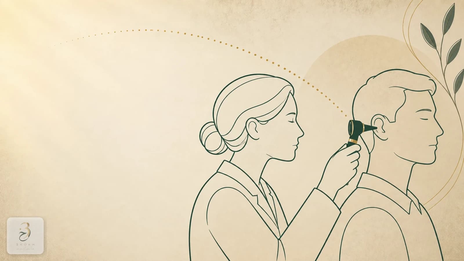
Otoscopy is a direct visual examination of the ear canal and the visible surface of the eardrum using a lighted magnifying instrument called an otoscope. It can reveal obstructing wax, canal inflammation or discharge, a foreign body, and changes in the eardrum, but it does not measure hearing or assess the inner ear by itself.
It is often the first part of an assessment for pain, itching, blockage, discharge, tinnitus, or hearing change. Findings must be interpreted alongside symptoms, history, and any tests that follow. This guide provides general education rather than an individual diagnosis. See the clinic's otoscopy service page for practical appointment information.
How is an otoscopy exam performed?
The examination is usually done while you are seated and requires no fasting, anaesthesia, or special preparation. The clinician first inspects the outer ear, selects a clean or disposable speculum that fits the canal, gently moves the pinna to straighten the view, and advances the tip only into the entrance of the canal.
The otoscope's light and magnification reveal the canal walls and eardrum. A pneumatic otoscope may also apply a very gentle change in pressure to assess eardrum movement. The MedlinePlus ear examination guide, reviewed in 2025, describes this process and confirms that routine preparation is not required.

Does otoscopy hurt?
Gentle otoscopy in a non-inflamed ear is usually not painful and may feel only like light touch or pressure. If the canal is infected, swollen, or very tender, moving the outer ear or touching it with the speculum can be uncomfortable or painful; sharp or increasing pain is not something to push through and should be reported immediately.
A correctly sized speculum, a clinician's hand braced against the face, and keeping still briefly reduce discomfort. Young children may sit securely on a caregiver's lap to prevent sudden movement. MedlinePlus notes that infection can make the examination uncomfortable and that the exam should stop if pain worsens.
What does the clinician inspect?
Otoscopy covers two main areas: the external ear canal and the eardrum. The clinician looks for obstructing wax, swelling, redness, discharge, a foreign body, or narrowing, then assesses the eardrum's colour, transparency, position, integrity, andΓÇöin pneumatic otoscopyΓÇömobility.
| Otoscopy finding | What it may suggest | Important limitation |
|---|---|---|
| Wax blocks the view | Cerumen accumulation or impaction | The eardrum cannot be judged until it is safely visible |
| Swollen, tender canal or discharge | External-ear inflammation or infection | Appearance alone does not identify the organism |
| Bulging or poorly mobile eardrum | Possible middle-ear fluid or inflammation | Symptoms and sometimes tympanometry are also needed |
| Visible hole in the eardrum | Eardrum perforation | Size and hearing effect need further assessment |
| Normal-looking canal and eardrum | No obvious outer-ear or eardrum abnormality | It does not prove normal hearing or a healthy inner ear |
This table helps explain report language; it is not a self-diagnosis tool. The American Academy of Pediatrics guideline describes eardrum contour, colour, translucency, and mobility as findings that must be interpreted with the child's symptoms.
What does an otoscopy result mean?
An otoscopy result is not simply ΓÇ£positiveΓÇ¥ or ΓÇ£negativeΓÇ¥; it records what was visible and how complete the view was. The examination may be normal, show an obvious finding such as wax or a foreign body, or remain limited because wax, discharge, or a narrow canal obscures the eardrum.
Redness alone does not equal infection, and a normal appearance does not exclude every cause of pain or hearing loss. Diagnosing a middle-ear infection depends on history and findings such as eardrum bulging or middle-ear fluid. The CDC likewise describes diagnosis as a combination of symptom assessment and examination of the eardrum. A useful follow-up question is: ΓÇ£What was seen, what remains uncertain, and what is the next step?ΓÇ¥
What can otoscopy not show?
Otoscopy shows the canal and eardrum surface, but it does not measure hearing thresholds, classify hearing loss, test the auditory nerve, or rule out all inner-ear disorders. A person can therefore have genuine hearing loss while the eardrum looks normal.
Tympanometry evaluates eardrum movement and middle-ear function by changing pressure, while audiometry measures the ability to hear sounds at different frequencies and levels. The NHS hearing-test guide explains these as separate tests. The next test should be chosen for the symptom and examination finding rather than applied as the same package to everyone.
Can earwax affect the result?
A large amount of wax can partly or completely block the view of the eardrum; ΓÇ£eardrum not visualisedΓÇ¥ is therefore not a valid conclusion about the eardrum itself. The clinician decides how and when to remove wax after considering the canal, previous perforation or ear surgery, and relevant medical factors.
Do not prepare for the appointment by inserting cotton buds, pins, or home tools, because they may push wax deeper or injure the canal and eardrum. ENT Health from the American Academy of OtolaryngologyΓÇôHead and Neck Surgery explains that wax needs attention when it causes symptoms or prevents a necessary examination. If you have had a perforation, ventilation tube, or ear surgery, ask a clinician before using drops or home irrigation.
What should parents know about a child's ear exam?
The examination principle is the same in children, but head support and speculum size must suit the child's age. A young child may sit on a caregiver's lap so the head and hands remain still for a few seconds; this is intended to prevent sudden movement, not to force a painful examination.
Explain simply that the clinician will look into the ear with a small light. Mention tenderness before the ear is touched. Crying and movement can limit the view and may make some visual changes harder to interpret, so repeat otoscopy or tympanometry may occasionally be needed. Fever, ear pulling, or redness alone is not enough to establish a middle-ear infection.
When is otoscopy not enough, and when is care urgent?
Otoscopy is not enough when the question concerns hearing performance or the inner ear. Gradual hearing loss, one-sided tinnitus, dizziness, recurrent infections, or persistent blockage after treatment may call for audiometry, tympanometry, or an ENT assessment.
Do not assume sudden hearing loss in one or both ears is wax; seek urgent medical assessment. The US National Institute on Deafness and Other Communication Disorders treats sudden sensorineural hearing loss as a medical emergency. Bloody or pus-like discharge, swelling around the ear, high fever, severe dizziness, facial weakness, or marked illness also needs prompt evaluation. Otoscopy is a useful first step, but it must not delay care for warning signs.
Sources
Frequently asked questions
Is otoscopy painful?
A gentle exam in a non-inflamed ear is usually not painful and may feel like light touch or pressure. Infection, swelling, or canal tenderness can cause discomfort; tell the clinician immediately if pain is sharp or increasing.
Do I need to prepare for otoscopy?
Usually not; fasting and anaesthesia are unnecessary. Do not insert anything into the ear before the visit, and mention a previous perforation, ventilation tube, ear surgery, or current ear drops.
Can otoscopy diagnose an ear infection with certainty?
Otoscopy can show important findings such as canal swelling, discharge, eardrum bulging, or reduced mobility, but diagnosis also uses symptoms and history. Tympanometry or another assessment may sometimes be needed.
Does a normal otoscopy result mean my hearing is normal?
No. Otoscopy checks the canal and eardrum appearance; it does not measure hearing thresholds or inner-ear function. Audiometry may still be needed for a hearing complaint even when the eardrum looks normal.
Can earwax prevent the eardrum from being seen?
Yes. Heavy wax may cover some or all of the eardrum, so the clinician decides how to remove it safely and when to repeat the examination. Cotton buds can push wax farther inward.
Which ear symptoms need urgent assessment?
Sudden hearing loss, bloody or pus-like discharge, swelling around the ear, high fever, severe dizziness, facial weakness, or marked illness should be assessed promptly rather than assumed to be wax.
Comments
No comments yet. Be the first to write one.